
Melissa
Kaplan's
Herp Care Collection
Last updated
January 1, 2014
Inclusion Body Disease in Boa Constrictor
Case II-165 or 96N165 (AFIP 2548995)
Armed
Forces Institute of Pathology Conference,
September 11, 1996
|
|
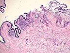
Necrotizing enteritis and ulceration in a red-tailed boa constrictor with zygomycosis and concurrent boid inclusion disease. (HE, 40X, 63K)
|
|
|
Pleomorphic fungal hyphae within the ulcerated areas of the enteric mucosa which have non-parallel walls, range up to 20 microns in diameter, are pauciseptate, and stain well with HE - all characteristics of a zygomycete. Further speciation is not possible based on examination of this fungus in tissue section. (HE, 400X, 50K) |
|
|
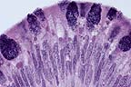
Numerous round cytoplasmic inclusions with enteric mucosal epithelial cells, which are characteristic findings in boid inclusion disease. Inclusions were also seen in lymphocytes and cells of the myenteric plexus. (HE, 400X, 32K) |

Signalment:
3-year-old
red-tailed boa constrictor (Boa constrictor).
History:
This boa was part of a colony of over 40 exotic snakes of various
species. It had a 5 month history of lethargy, intermittent anorexia,
weight loss, and fetid loose feces. On physical examination it had dehydration
and petechiation of its oral mucosa and ventral scutes. It was treated
with fluids and antibiotics over a period of 2 months without improvement.
Gross
Pathology:
Marked coelomic and pericardial serous effusions. Diffuse petechiation
of fat bodies and subcutis. Focal segment of distal small intestine with
severe edema and luminal accumulation of partially adherent thick mucoid
material.
Laboratory Results:
- CBC ( 4 weeks ante-mortem): PCV = 21%, WBC = 1500 cells/ul
- Intestinal contents, fungal culture: Small numbers ofTrichosporon beigelii, Aspergillus clavatus; Salmonella culture negative
- fecal floatation negative
- IFA for Giardia and Cryptosporidium negative.
Contributor's
Diagnosis and Comments:
Necrotizing enteritis, heterophilic and granulomatous, moderate,
focal, with intra-lesional fungal hyphae and yeast forms - presumptive
etiology: Aspergillus clavatus, Trichosporon beigelii.
Intra-cytoplasmic eosinophilic inclusion bodies in most cell types - Boid inclusion body disease.
Inclusion body disease (IBD) affects various species of boid snakes and usually manifests as progressive debilitation, anorexia, weight loss, regurgitation, and neurologic signs. The presence of the typical, well-defined intracytoplasmic eosinophilic inclusion bodies is often associated with cellular degeneration in various tissues, most commonly in the CNS, where a non-suppurative encephalomyelitis can also be observed. Various secondary infections are frequently associated with IBD, such as pneumonia and nephritis. In this boa, encephalitis was not noted, although inclusions were numerous in the brain, and were associated with degeneration. The animal also had a mycotic enteritis affecting a focal segment of distal small intestine. The morphology of the fungal elements (pleomorphic hyphae, yeast forms) was compatible with both the Aspergillus clavatus and Trichosporon beigelii organisms obtained by fungal culture of the intestinal contents. The boa probably developed fungal enteritis from the changes in gut flora induced by prolonged antibiotic treatment; IBD may have been a predisposing factor. Electron microscopy of liver tissue revealed that the inclusions contained granular homogenous osmiophilic material. Lined around the periphery, and occasionally inside the inclusions were numerous clathrin-coated pinocytotic vesicles. A few particles consistent with type C retroviruses, measuring 95-110 nm, were observed in the intercellular spaces, sometimes budding from the cell membrane. The particles were similar to those previously reported in snakes with IBD (see reference).
AFIP
Diagnosis:
Intestine: Enteritis, ulcerative, necrotizing, granulomatous, multifocal,
severe, with fungal hyphae, red-tailed boa constrictor (Boa constrictor),
reptile, etiology consistent with a zygomycete.
Intestinal epithelium; lymphocytes; intestinal ganglion cells of myenteric plexi: Eosinophilic intracytoplasmic inclusion bodies.
Conference
Note:
The inclusions in submitted tissues are eosinophilic to amphophilic,
range up to 10 microns in diameter, and did not appear to be associated
with degenerative or inflammatory changes. The morphologic features of
the fungus are consistent with the those of the Class Zygomycetes and
not with Aspergillus or Trichosporon spp. These features
include broad, thin-walled, infrequently septate, pleomorphic hyphae that
range from about 5 to 20 m in width and irrgeular right angle branches.
The fact that the hyphae stain well with H&E is also characteristic
of a zygomycete.
Inclusion body disease of boid snakes has been recognized for over 20 years in private and zoological collections in the United States. The disease affects only snakes of the Family Boidae including both boa constrictors and pythons. The disease is characterized by the formation of intracytoplasmic inclusions in the epithelial cells of all major organs and neurons in the central nervous system. Clinical symptoms include head tremors, disorientation, incoordination, and regurgitation. Ultrastructurally, a type C retrovirus has been associated with the lesions. The presence of a retrovirus is supported by demonstration of reverse transcriptase activity in the plasma and within the supernatant from primary kidney cell cultures of affected snakes.
Major histologic lesions include a nonsuppurative meningoencephalitis with neuronal degeneration, gliosis, and demyelinization. Intracytoplasmic inclusions are noted within degenerating neurons of the gray matter and ependymal cells. Inflammation in the central nervous system is more severe in pythons; however, intracytoplasmic inclusions are more numerous in boa constrictors. In the experience of pathologists at the National Zoological Park, Washington D.C., inclusion body disease has been found not only as a primary disease but also in association with secondary infections or as an incidental finding. In fact, many lesions within visceral organs of affected snakes have been attributed to secondary infections.
Contributor:
Dept of Veterinary Pathology, Microbiology & Immunology, School
of Veterinary Medicine, University of California, Davis, CA 95616.
Reference:
Schumacher J, Jacobson ER, Homer BL, Gaskin JM. Inclusion body disease
in boid snakes, J Zoo and Wildlife Med 25(4):511-24, 1994.
Related Articles