
Melissa
Kaplan's
Herp Care Collection
Last updated
January 1, 2014
Identification and treatment of metabolic bone disease
©1995, 2002 Melissa Kaplan
|
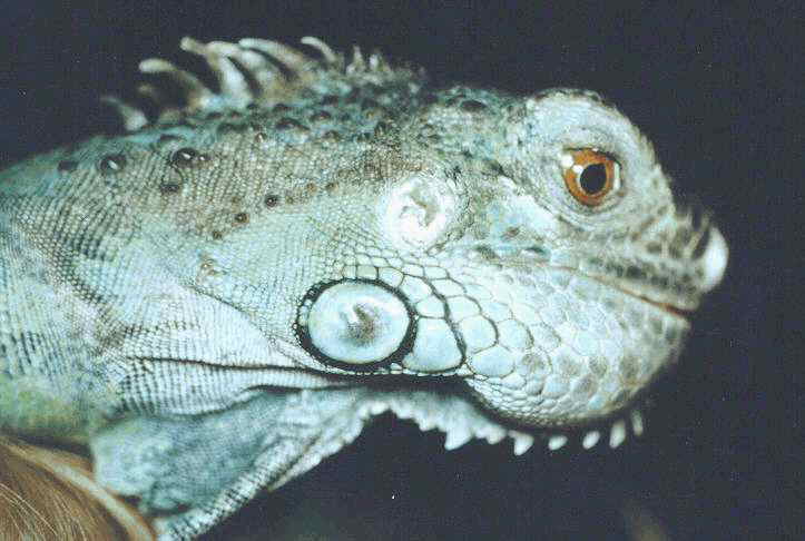
Rugwort at 3 years of age. He came to me in 1994 at age 20 months, already severely deformed and almost paralyzed by severe MBD. To this day, his skull, spine, and hip joints are still deformed from the MBD and malnutrition caused by the lousy environment and lettuce-and-cat-food diet provided by his first owners who couldn't afford to care for him properly yet took almost a year to finally give him to someone who could.* |
Metabolic bone disease (MBD) is an umbrella term that covers a number of disorders related to the weakening of the bone or impaired systems function caused by an imbalance in vitamin D3, calcium and phosphorus. This imbalance may be caused by a lack of or too much of one of these three essential elements or the failure to provide one or more of them in a bioavailable form. Many foods highly touted for their calcium content, such as spinach, carrots, collards, chards and other thick leafy greens, contain calcium oxalates that bind calcium. This renders most or all dietary calcium, both that contained in the foods and that added to the foods as supplements, unavailable for maintenance and growth, depending on the quantities ingested. MBD and calcium metabolism is discussed in great detail in many texts and so will not be elaborated upon here. |
Quite simply stated, vitamin D3 (dietary or derived from exposure to ultraviolet B), calcium (dietary and matter recycled from the bone matrix) and phosphorus (dietary) interact together to perform a number of functions besides bone growth and maintenance, including muscle contractions and blood coagulation. The result is a well-functioning system, with calcium restored to and, in the case of growing animals, added to the bone matrix. Too much phosphorus can throw this process off, as can too much or too little vitamin D3 or too little access to ultraviolet B wavelengths. As the dangers of calcium deficiency become more widely known, there is increased risk that pet owners may add too much calcium to their reptile's diet. This results in hypercalcemia, a condition as fraught with peril as is hypocalcemia. To date, however, hypercalcemia is quite rare, occurring most often in healthy gravid iguanas in which such a state is desirable for the health of the female and the developing embryos (see the article on dystocia).
Signs of metabolic bone disease include hard knobs in the long bones of the legs, bumps along the vertebral column of the back and tail, bilateral softening or hard swelling of the lower jaw, and softening of the plastron or carapace. All of these signs may be felt before they can be seen, making a careful physical exam important. Visible signs of moderate to severe MBD include jerky gait when walking, repeated tremors and twitches in the limbs and muscles of the legs and toes when at rest, and shakiness when being held. (The occasional single myoclonic jerk that happens is not considered indicative of MBD.)
Advanced cases of MBD include all the above signs plus constipation, anorexia and fractured bones. Severely deficient reptiles tend to be lethargic and may only be able to drag themselves along the ground. Arboreal lizards spend all of their time on the ground as they lack the strength to grip and climb.
Moderate to severe cases of MBD require the proper diet, temperatures, and light wavelengths as well as a more powerful calcium supplement than those found in pet stores. Oral administration of calcium glubionate (NeoCalglucon®, 1cc/kg PO bid prn) or injections of calcium lactate (Calphosan®, 250 mg/kg IV/IM, bid) or calcium gluconate (100 mg/kg IM qid prn) are generally prescribed by veterinarians. Mader (1993) reports faster recovery with calcitonin (Calcimar®, Miacalcin®, 50 IU/kg IM in front leg, repeated once a week for two weeks) when it is administered to iguanas who have been returned to normal serum calcium levels. Use of calcitonin before normal levels have been established, however, may cause hypocalcemic tetany and death. Mild cases, cases where the signs are felt or just barely visible, may successfully be treated by providing the proper environment and diet. In the case of diurnal lizards and chelonians, proper environment includes not only the proper temperature ranges and diet, but daily access to ultraviolet B wavelengths.
There remains much debate as to the necessity of and efficacy of natural and artificially produced ultraviolet B wavelengths in the development of precursors to vitamin D3 and the metabolism of calcium. Gehrmann (1991) reports anecdotally that not all lizards require UVB to maintain proper D3-calcium-phosphorus balance. Bernard, et al. (1991) found that the green iguana (Iguana iguana) fared much better when exposed to ultraviolet B wavelengths than they did to vitamin D3 injections or supplements added to their food. As both ultraviolet A, which acts upon appetite and behavior, and ultraviolet B wavelengths are more likely to benefit lizards and chelonians than not, long daily periods of access to such wavelengths should be considered a necessary part of the care of diurnal lizards and all chelonians (Alberts, 1994), and should figure as a prominent part in the daily care and maintenance of reptiles who commonly suffer from MBD in captivity, such as the green iguana.
Along with proper day and night temperature gradients and a nighttime dark period of sufficient length (based on native habitat), proper diet is essential to recovery. Herbivores and omnivores should be fed calcium-rich, nutrient dense foods such as squashes, green beans, alfalfa (from alfalfa powder, crushed alfalfa tablets, alfalfa tea, or softened rabbit food pellets or pulverized hay cubes), parsnips, mustard greens, dandelions, escarole, and fruits such as figs, papaya, cantaloupe and berries (Barten, 1993; Frye, 1991). The food should be supplemented with additional calcium and a multivitamin formulated for reptiles or birds, or a crushed Centrum vitamin tablet formulated for humans (Donoghue, 1996). Omnivores and carnivores should be fed whole captive bred prey (to reduce the risk of zoonotic infection from parasites commonly found in wild prey) that have been raised on nutritious foods or have been gut loaded with nutritious foods for several days before being fed out.
The references included above may be found in my iguana bibliography.
* Rugwort died, of kidney failure, just after Christmas, 2001.
Related Articles